Activity #3: Onion and Cheek Cell Analysis

Materials:  foldscope kits, white onions, iodine, toothpicks, methylene blue

Onion Cell Analysis

Part 1: Click here and carefully view the to learn how to view onion cells using a microscope. Use a few drops of iodine to help visualize the different parts of the onion cells. Describe the appearance of your onion cells in your workbook. 

Part 2: Take a picture of your onion cells and share it below. Take time to view other team submissions. Enter the first name of each team member in the “Subject” field . A QR code is provided below for ease of upload. 

Made with Padlet
Cheek Cell Analysis

Part 3: Each student should gently scrape the inside of their cheek with a clean toothpick to avoid discomfort. Transfer the cells by stirring the toothpick in a drop of methylene blue placed in the center of a clean microscope slide. This stain will highlight the cells’ nuclei and other internal structures, making them easier to observe. Place a cover slip over the sample. Refer to the video below for additional instructions. Apply the techniques demonstrated in the video to your provided Foldscope materials.

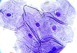
Part 4: Observe the structure of their cheek cells, noting the cell membrane (outside), cytoplasm (inside), and nucleus (dark portion in the middle that contains the DNA). Describe the appearance of your onion cells in your workbook. See image below as a reference. 

Part 5: Take a picture of your cheek cells and share it below. Take time to view other team submissions. Enter the first name of each team member in the “Subject” field . A QR code is provided below for ease of upload. 

Made with Padlet